Fig 7.
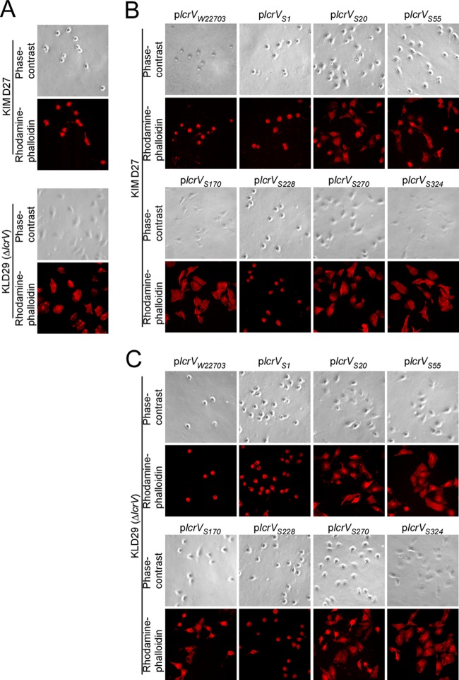
Strep-tagged LcrV and Yersinia pestis effector translocation. (A) Y. pestis strains KIM D27 (wild-type lcrV) and KLD29 (ΔlcrV) were used to infect HeLa tissue culture cells for 3 h at an MOI of 10. Samples were fixed, stained with rhodamine-phalloidin, and imaged by fluorescence or phase-contrast microscopy to reveal actin cable rearrangements and cell rounding as a measure for effector translocation. (B) Y. pestis KIM D27 strains harboring lcrV plasmids (plcrVW22703, plcrVS1, plcrVS20, plcrVS55, plcrVS170, plcrVS228, plcrVS270, and plcrVS324) were subjected to the same assay as described for panel A. (C) Y. pestis KLD29 strains harboring lcrV plasmids (plcrVW22703, plcrVS1, plcrVS20, plcrVS55, plcrVS170, plcrVS228, plcrVS270, and plcrVS324) were subjected to the same assay as described for panel A.

