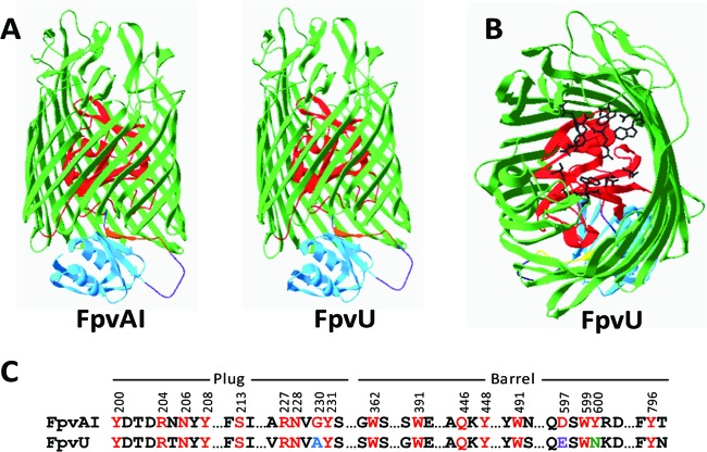
Fig 1.
(A) Homology model of FpvU from P. protegens Pf-5 compared to FpvAI from P. aeruginosa PAO1 showing the structural components, with the β barrel in green, plug in red, N-terminal signaling domain in blue, connecting loop in purple, and TonB box in brown. (B) Positions of amino acid residues in the plug and β-barrel domains of FpvU corresponding to the residues of FpvAI that are involved in pyoverdine binding. Amino acid side chain structures are shown in black. (C) Alignment of FpvAI and FpvU. Amino acid residues of FpvAI that interact with the PAO1 pyoverdine (8, 13) are numbered and shown in red font. Identical residues in FpvU are in red, conservative substitutions are in blue, semiconservative substitutions are in purple, and nonconservative substitutions are in green.

