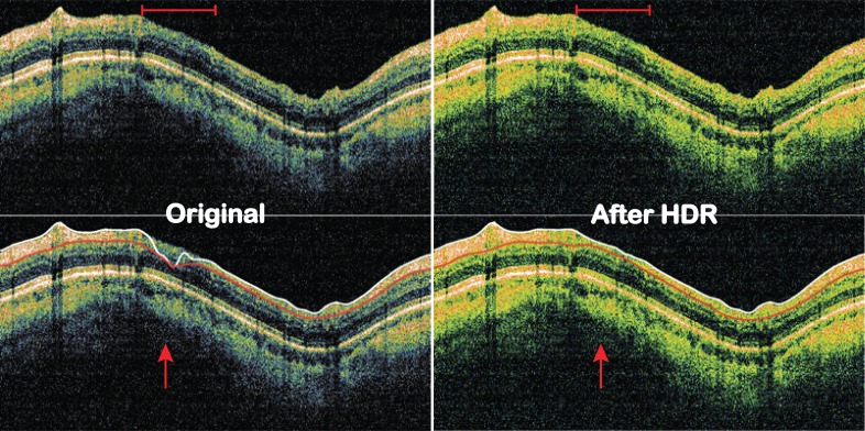
Figure 2. .
SD-OCT images before and after HDR imaging. Top row: visibility of the retinal layers became clearer across the image, especially the area within the red bar on top. Signal levels also became more homogeneous with HDR imaging. Bottom row: RNFL segmentation failed on original image but succeeded after HDR imaging (red arrow).

