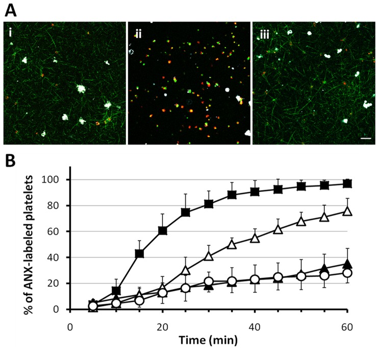
Figure 5. Attenuation of fibrin network formation suppress PS exposure on platelets surface.
(A) Exposure of PS by R-6G labeled platelets (red) in the presence of either (i) FK633 (30 µM), (ii) GPRP (3 mM) or (iii) Cyt-B (100 µg/ml) upon stimulation of diluted PRP containing fbg-488 (green) and ANX (white) with thrombin (1 U/ml). CLSM images were taken 60 minutes after thrombin supplementation. (B) Kinetics of platelet PS exposure expressed as the percentage of ANX fluorescence-positive platelets in thrombin-treated (1 U/ml) samples in CLSM study. Presence of either FK633 (open triangle, n = 7), GPRP (open circle, n = 7) or Cyt-B (closed triangle, n = 7) suppressed platelet anionic phospholipids exposure compared with control (close square, n = 5), suggesting that crosstalk between platelets and the fibrin scaffold is a key feature of the anionic phospholipids exposure. Data are shown as mean ± SD. Scale bar shows 10 µm.

