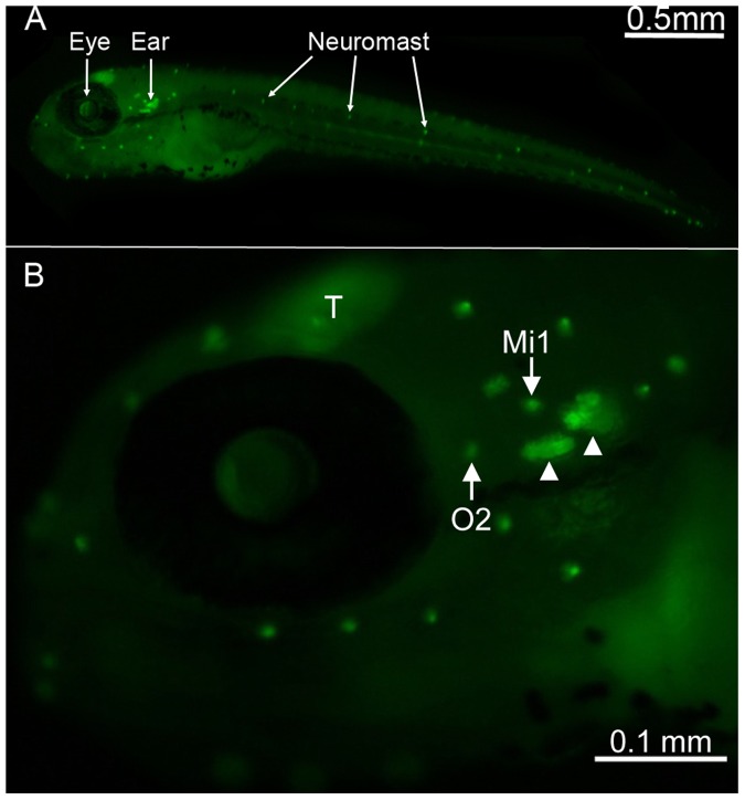
Figure 1. Location of neuromasts on Brn3c-GFP transgenic zebrafish.
(A) Lateral view of a 5 days post-fertilization transgenic zebrafish showing GFP expression in neuromasts that are found along the head and the body (bright dots) of the animal. (B) Higher magnification of the head region, with neuromasts containing brightly GFP-labeled hair cells (white arrows) and inner ear organs (white arrowheads) easily identifiable. The otic 2 (O2) and middle 1 (Mi1) neuromasts are highlighted as these are the two neuromasts from which data for this study were obtained. The zebrafish optic tectum (T) is another structure that is also labeled with GFP.

