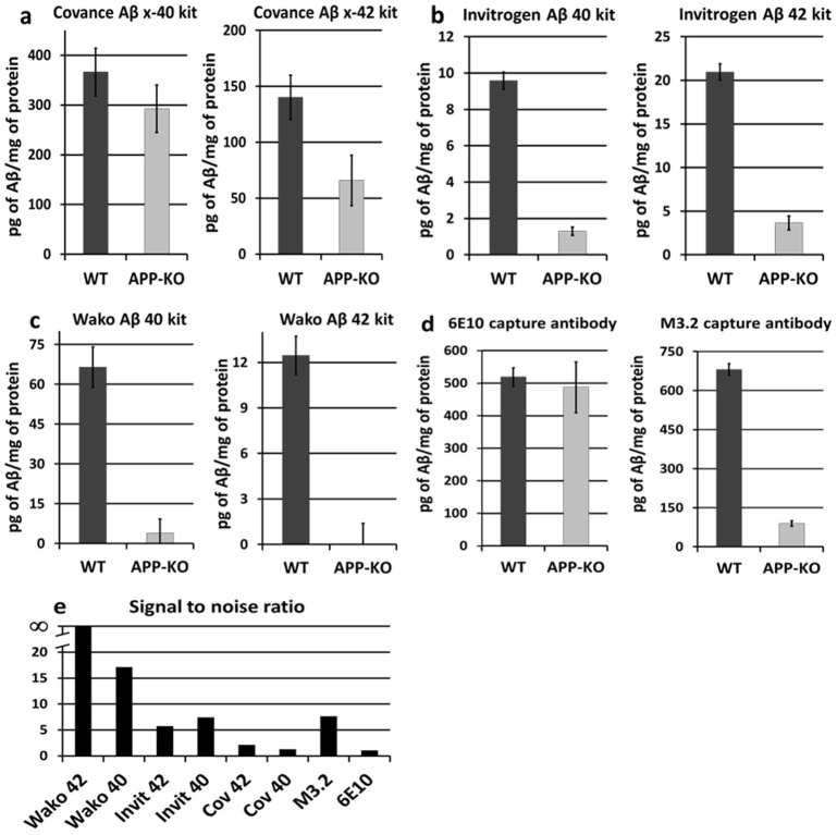
Figure 1. The amount of background signal with APP-KO tissue varies widely between different ELISAs.
A) The Covance Colorimetric BetaMark™ Beta-Amyloid x-40 ELISA Kit (left) and x-42 Kit (right). Both kits give a signal with APP-KO tissue that is significantly above baseline. The x-40 kit gives a signal with APP-KO tissue that is not significantly different from WT (the difference between APP-KO and WT is significant for the x-42 kit). B) The Invitrogen Aβ 40 Mouse ELISA Kit (left) and Aβ 42 Mouse ELISA Kit (right). Both kits give a signal with APP-KO tissue that is significantly above baseline, but also significantly different from WT tissue. C) The Wako β-Amyloid (40) ELISA Kit (left) and β-Amyloid (42) ELISA High-Sensitive Kit (right). Both kits give a signal with APP-KO tissue that is not statistically significant from baseline. D) Both WT and APP-KO give a similar level of background signal with an ELISA for human β-amyloid (6E10 capture antibody – left), but show a clear difference when a rodent-specific capture antibody is used (M3.2 antibody – right). E) “Signal to noise” ratio of the WT signal divided by the APP-KO signal for each ELISA. Error bars in A–D are standard error.

