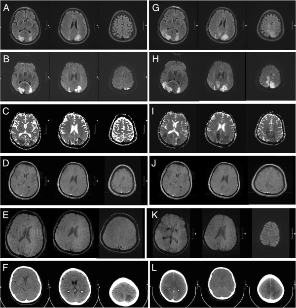
Figure 1.
Serial magnetic resonance imaging (3 Tesla) and computed tomography scans showing progressive bilateral reversible posterior leukoencephalopathy syndrome. (A–E) Magnetic resonance imaging immediately after the patient was admitted showed (A) marked hyperintensity in T2 and fluid-attenuated inversion recovery (FLAIR) sequences of the posterior lesions. Diffusion-weighted imaging exhibited restricted diffusion (B) with a decreased signal on apparent diffusion coefficient mapping (C) consistent with cytotoxic edema. Lesions showed slight contrast enhancement (D). Susceptibility-weighted imaging (SWI) showed signal loss indicating the beginning of hemorrhagic lesion transformation (E). (F–J) Magnetic resonance imaging two days after the patient was admitted showed progressive confluent lesions in T2 and FLAIR sequences (F). Increasing areas of restricted diffusion (G) and volume expansion (H) indicated progressive cytotoxic edema. Lesions were still slightly contrast enhancing (I). SWI showed progressive signal loss indicating advancing hemorrhagic lesion transformation (J). (K) A computed tomography scan immediately after admission demonstrates well-demarcated bilateral hypodensities in posterior brain regions. (L) The computed tomography scan on day 3 after admission reveals global cerebral swelling, subsequent upper and lower brainstem herniation and brainstem compression as well as generalized thrombosis of the cerebral sinus and veins.

