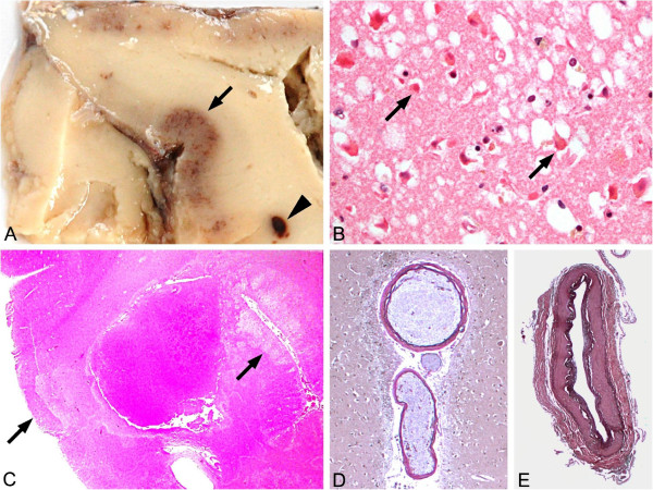
Figure 2.
Neuropathological analysis revealing laminar necrosis and hypoxic neuronal cell death in the absence of overt arterial pathology. (A,B) Gross appearance revealed cortical discoloration due to laminar necrosis (→) and small white matter hemorrhage (►) in occipitotemporal regions. (C) Histopathological analysis showed hypoxic neuronal damage (arrows) (that is, nerve cells with eosinophilic and shrunken cytoplasm and hyperchromatic condensed nucleus (hematoxylin and eosin stain ×100)) (D,E) but no evidence of any arterial vascular pathology except for minor arteriosclerosis of intracerebral (D) and basal vessels (E) (elastica-van Gieson stain ×100).

