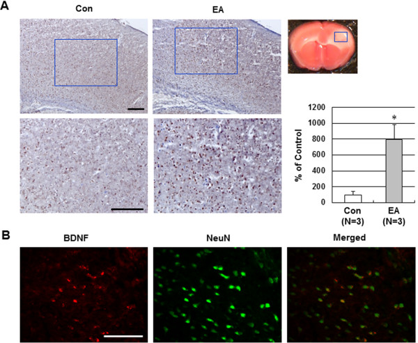
Figure 4.
Effect of EA preconditioning on BDNF expression in ischemic brain. (A) Representative immunohistochemical staining photographs showed BDNF-positive cell expression 24 h after occlusion in the ischemic cortex of mice that received control or EA. The blue rectangle represents the imaging field. Quantification of BDNF-positive cells is expressed as the % change of the control. The results are expressed as mean ± SEM for three mice in each group. *, P<0.05 vs. control (Con). (B) Representative double immunofluorescence staining for BDNF (red) and NeuN (neuronal marker, green) in EA-preconditioned ischemic brain. EA-induced BDNF expression was colocalized with the neurons after ischemic injury. Scale bar = 100 μm.

