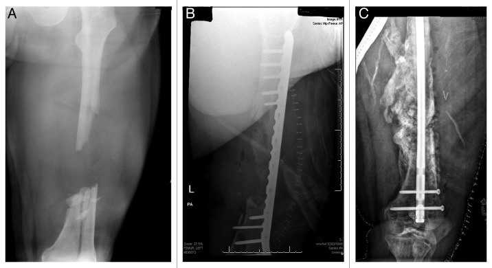
Figure 4:
(A) AP radiograph of a large, segmental femur fracture with a sizeable diaphyseal defect. (B) postoperative AP radiograph of the fracture following bridge plating and implantation of iliac crest bone graph, DBM and BMP. (C) AP radiograph taken ten months following injury showing expansive bone formation and subsequent hypertrophic nonunion following hardware failure, treated with rigid intramedullary fixation.

