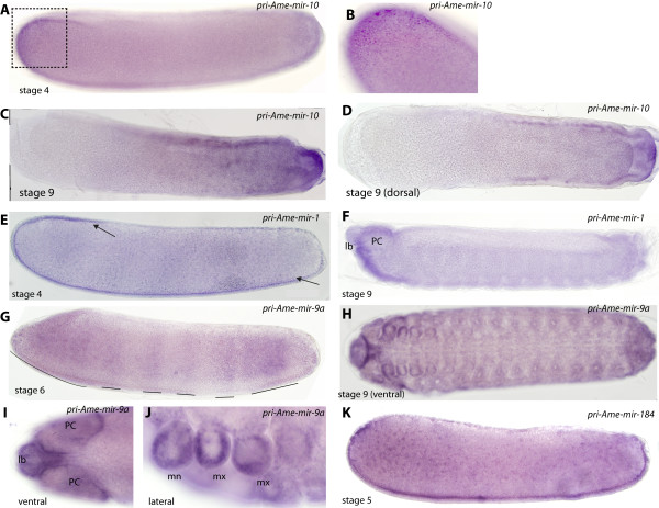
Figure 2.
Expression of honeybee pri-miRNAs present during embryogenesis. Embryos are shown with anterior to the left and dorsal side up, unless stated otherwise. Scale bar is 100 μm. (A) Expression of pri-Ame-mir-10 at stage 4 is detected in the ectoderm at the anterior pole of the embryo. (B) Higher magnification image showing nuclear staining in anterior ectodermal cells. (C and D) Expression of pri-Ame-10 at stage 9 is found in the posterior mesoderm (ventral orientation in D) and in the posterior terminal segment. (E) Pri-Ame-mir-1 is expressed central ventral ectoderm at stage 5. (F) By stage 9 pri-Ame-mir-1 RNA is detected in regions of the procephalic lobes (PC), and within the labrum (lb). Expression is also detected in the posterior anal plates. (G) Pri-Ame-mir-9a is detected in broad bands of ectodermal cells across the anterior-posterior axis. Staining was absent from the dorsal and ventral regions of the stage 6 embryo. (H) Expression of pri-Ame-mir-9a is detected through the epithelium at stage 9. (I) Strong epithelial staining for pri-Ame-mir-9a in the labrum and procephalic lobes at stage 9. (J) Pri-Ame-mir-9a is also present in the epithelium of the head appendages, maxillae (mx) and mandibles (mn) but absent from the internal regions of these structures, where the sensory neurons are present. (K) Expression of pri-Ame-mir-184 is detected through mesoderm from anterior, ventral and posterior of the embryo but absent from the ventral side of the stage 4 embryo.

