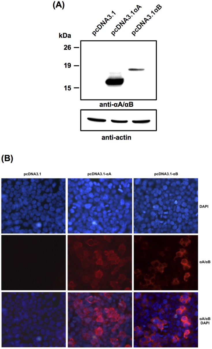
Figure 1. Expression of αA- and αB-crystallins in transiently transfected 293T cells.
(A) Western blot analysis of αA- and αB-crystallin levels 24 h post-transfection. Fifty micrograms of total proteins from cell extracts were subjected to 12% SDS-PAGE and immunoassayed with anti-αA/αB-crystallin to detect the overexpressed α-crystallins and with anti-ß-actin as a control of equal protein loading. (B) Immunofluorescence analysis with anti-αA/αB-crystallin showing cytoplasmic expression of αA- (pcDNA3.1-αA) and αB- (pcDNA3.1-αB) crystallins 24 h post-transfection, while no detection was observed in cells transfected with the empty plasmid (pcDNA3.1).

