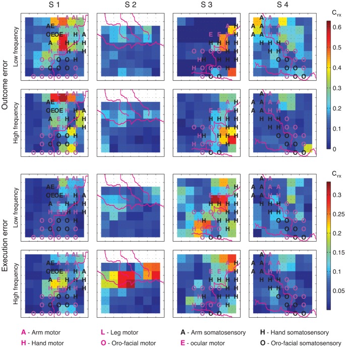
Figure 8. Spatial distribution of CYX for detection of outcome and execution errors.
Detection was performed by using either low or high frequency components of the signals recorded from electrode quartets (2×2 neighboring electrodes). Purple lines depict the central sulcus, the Sylvian fissure and, for S2 only, the pre and post central sulci. Letters in the squares mark the functional subarea (A – arm, H – hand, L – leg, E – ocular, O – oro-facial) in motor (purple) and somatosensory (black) cortex as determined by ESM. Every coloured square represents one quartet of electrodes with the electrodes at the corners of the square. Colours of the squares depict the normalized mutual information according to the colour bar. Since no recordings were made from the top row of grid electrodes for S2, we show the top row of quartets as white. The top left square in the ECoG grids correspond to the electrode closest to the red star in Figure 3. Detection was made using rLDA.

