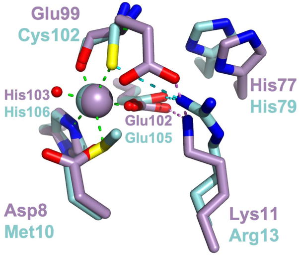
Figure 9.
Structural alignment of the primary site residues of IdeR•Co2+ (PDB ID 1FX7; carbons and Co2+ ion colored cyan) with the C site residues of E11K•Mn2+ (carbons and Mn2+ ion colored violet). Metal ligand interactions taking place between IdeR residues and bound Co2+ are shown as green dashed lines. Hydrogen bonding interactions taking place between Arg13 and primary site residues are shown as dotted cyan lines, while hydrogen bonds between Lys11 of E11K and C site residues are shown as dotted violet lines.

