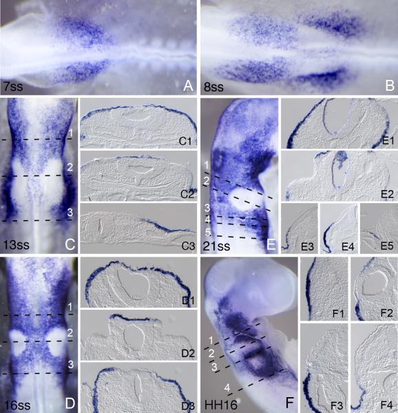
Figure 4.
Foxi2 expression in cranial ectoderm at different developmental stages. Numbers in each large panel refer to the embryonic stage in number of somites or by by Hamburger and Hamilton stage designated by HH. Embryos were processed for whole mount in situ hybridization and then sectioned transversely at 18μm. The level of each section is indicated by dashed lines. (A) Expression at 7ss when Foxi2 expression is first seen anterior to the otic placode and extending into the presumptive trigeminal region. (B) Expression at 8ss when second patch of Foxi2 expression is seen lateral to the otic placode and extending caudally to the first few pairs of somites. (C) Expression at 13ss. Foxi2 extends more anteriorly and posteriorly in cranial ectoderm with the striking exception of the thickened otic region. (D) Expression at 16ss, where the exclusion of Foxi2 from the otic placode is even more pronounced. (E) Expression at 21ss where Foxi2 expression extends more anteriorly and is still expressed strongly in the cranial ectoderm with the exception of the otic cup. (F) Expression at HH16 (equivalent to 26-28 pairs of somites). Expression is down regulated from the most of the cranial ectoderm. Expression is now restricted to the pharyngeal arches.

