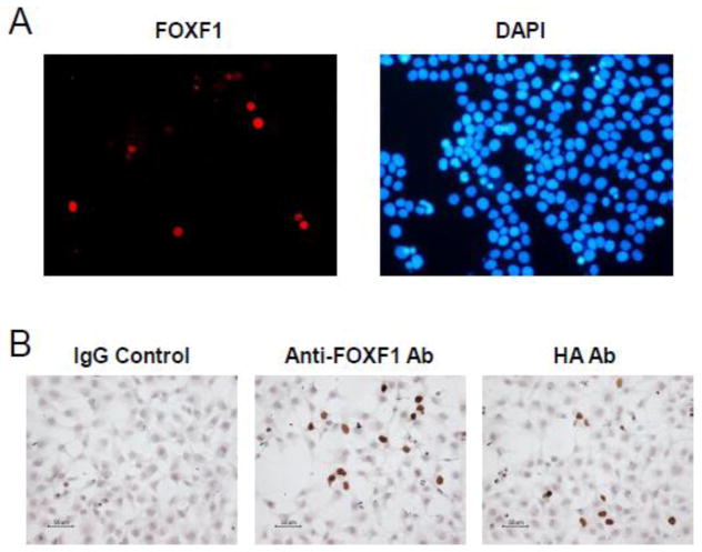
Fig. 1.
The anti-FOXF1 antibody specifically stains nuclear FOXF1 proteins in FOXF1-transfected colorectal cancer cells. (A) Immunofluorescence analysis of ectopic expression of HA-tagged FOXF1 in DLD-1 colorectal cancer cells using the anti-FOXF1 antibody. The left panel is HA-FOXF1-transfected DLD-1 cells stained with the anti-FOXF1 antibody and the goat anti-rabbit IgG Alexa 568 antibody. The right panel is the co-staining of cells with the DNA staining dye DAPI. (B) Immunocytochemistry analysis of HA-tagged FOXF1 expression in HCT116 colorectal cancer cells using either the anti-FOXF1 or anti-HA antibody. HA-FOXF1-transfected HCT116 cells stained with rabbit IgG and the anti-HA antibody served as negative and positive controls, respectively, in comparison with those stained with the anti-FOXF1 antibody. Immunocytochemistry analysis was performed as described in “Materials and Methods”.

