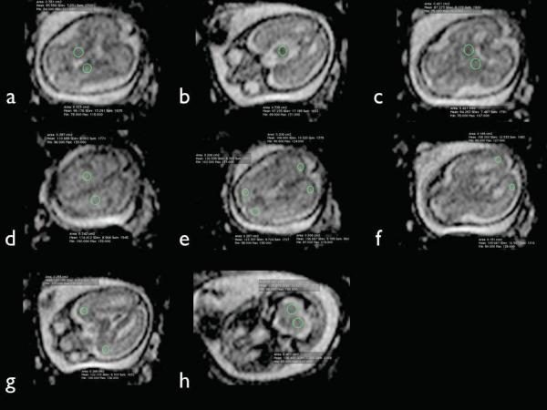
Figure 1.
Regions of interest (ROI) for fetal brain ADC measurements. Multiple axial ADC images from a fetal brain at 22.7 gestational weeks demonstrating sample ROI placement within the basal ganglia (a), pons (b), thalamus (c), centrum semiovale (d), frontal and parietal white matter (e), occipital white matter (f), temporal white matter (g), and cerebellar hemispheres (h).

