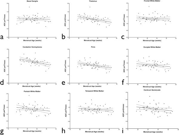
Figure 2.
Correlation between ADC values and menstrual age for different fetal brain regions: a) basal ganglia; R2=0.01; b) thalamus; R2=0.17; c) frontal white matter; R2=0.01; d) cerebellar hemispheres; R2=0.23; e) pons; R2=0.18; f) occipital white matter; R2=0.05, g) parietal white matter; R2=0.05, h) temporal white matter; R2=0.02, i) and centrum semiovale; R2=0.06. A significant menstrual agerelated decline in ADC values was observed in the pons, thalamus, and cerebellar hemispheres (p < 0.05).

