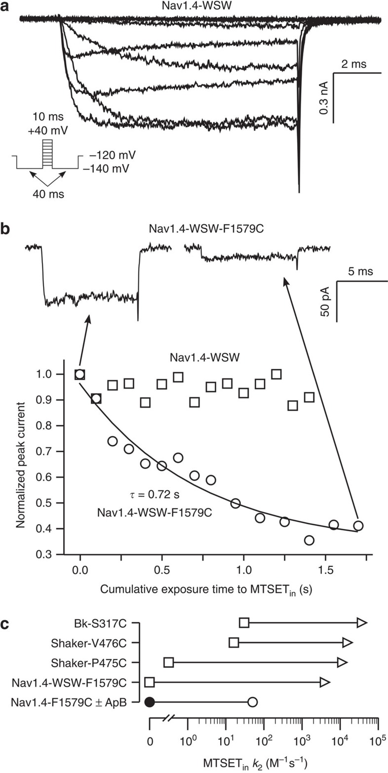
Figure 7. The open pore of Nav1.4-WSW is fully accessible to internal MTSET.
(a) Current responses to 10 ms voltage steps from −120 to 40 mV (20 mV steps) from a holding potential of −140 mV (see inset) from Nav1.4-WSW channels in inside–out patches excised from Xenopus oocytes. (b) The time course of the reduction in peak current for Nav1.4-WSW and Nav1.4-WSW-F1579C with respect to the cumulative exposure time to internal MTSET at 0 mV (see methods). Current responses for Nav1.4-WSW-F1579C before and after 1.7 s of exposure to internal MTSET at 0 mV are shown. (c) Second-order reaction rates of internal MTSET with Nav1.4-WSW-F1579C in the closed (square) and open (triangle) states. The reported change in closed to open state reaction rates for several residues in the pore of Shaker and BK potassium channels are shown for comparison41,42, as well as the reaction rate for Nav1.4-F1579C without (closed circle) and in the presence of the fast inactivation disrupting toxin Anthopleurin B (ApB) (open circle)40.

