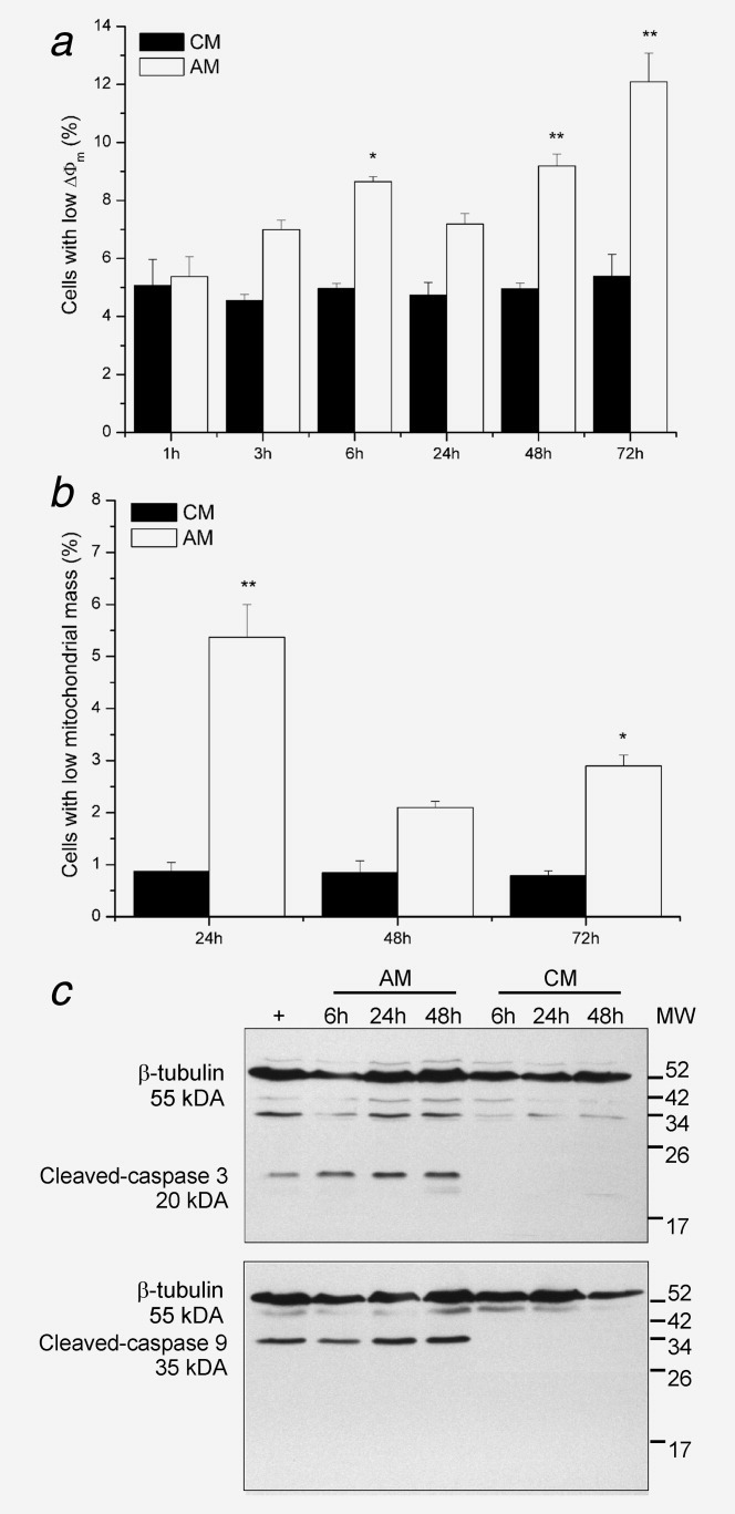
Figure 4.

AM activates the mitochondrial pathway of apoptosis in PC-3 cells. (a) Percentage of cells with low ΔΨm as measured by JC-1. *p < 0.05, **p < 0.001 (ANOVA). (b) Percentage of cells with decreased mitochondrial mass as measured by NAO. *p < 0.001 (ANOVA). (c) Western-blot analysis of protein lysates from cells treated with AM or CM. + Lysate from Jurkat cells treated with 25 μM etoposide. Data are presented as mean ± SEM of three independent experiments (panels a and b) or representative of three independent experiments (panel c).
