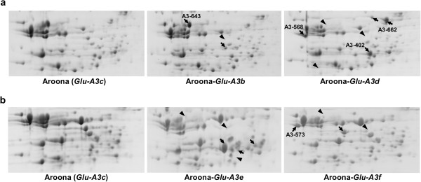
Figure 3.

Separation of flour proteins in Glu-A3 NILs using two-dimensional gel electrophoresis (2D-PAGE). a) Comparison of storage proteins from Aroona, Aroona-Glu-A3b and Aroona-Glu-A3d. b) Comparison of storage proteins from Aroona, Aroona-Glu-A3e and Aroona-Glu-A3f. The arrows indicate the unique protein spots in each NIL, and the arrowheads show the protein spots present in Aroona but absent in other NILs. The high molecular weight glutenin subunit protein spots are the same and not shown here due to limited space.
