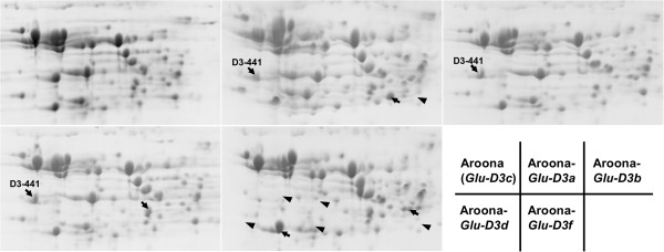
Figure 5.

Separation of flour proteins in five Glu-D3 NILs using two-dimensional gel electrophoresis (2D-PAGE). The arrows indicate the unique protein spots in each NIL, and the arrowheads show the protein spots present in Aroona but absent in other NILs.
