Figure 1.

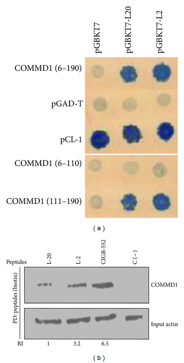
Peptides and COMMD1 interact in mammalian cells. (a) Yeast two hybrids showing interactions between full length COMMD1 and COMMD1 (111–190) prey fusions and the indicated L-2 and L-20 peptides as baits tested by interaction mating using SD Leu/Trp/His/Ade plates. pCL1 was used as a positive control of growth and the interaction between empty bait vector pGBKT7 and empty prey pGAD-T was used as a negative control. Interactions between pGBKT7 and preys constructions and pGAD-T and baits were used as a control of the interaction specificity. (b) Pull-down assay demonstrating specific COMMD1/peptides complex in H460 cells. Endogenous COMMD1 was precipitated from H460 cell lysates. The biotinylated peptides bond to streptavidin sepharose was used as bait and cell lysates as prey. The precipitated material was analysis by Western blot using antibodies directed against endogenous COMMD1. C (−) indicates streptavidin sepharose without peptide. Actin was used as input housekeeping. Relative levels of precipitated COMMD1 were determined by densitometry and normalized to actin. Ratios were depicted under each lane (RI) relative to COMMD1 levels immuprecipitated with peptide L-20. ImajeJ was used for quantification.
