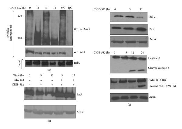
Figure 3.

CIGB-552 induces ubiquitination and proteasomal degradation of RelA. (a) H460 cells were treated with CIGB-552 (25 μM) for the times specified or MG132 (25 μmol/L) for 5 h. Immunoprecipitation (IP) of ubiquitinated protein following antiubiquitin Western blot analysis (WB) show an increase of ubiquitinated forms of RelA 2 h after CIGB-552 treatment. The control of immunoprecipitation was done with anti-rabbit IgG. RelA in input samples is shown. MG indicates MG132 (positive control). (b) H460 cells were treated with either CIGB-552 (25 μM) and MG132 (25 μmol/L) for the indicated times. Anti-RelA immunoblot shows native protein in whole-cell extracts. The levels of RelA were increased in cells treated with CIGB-552 and MG132 with respect to the cells treated alone with CIGB-552, indicative of proteasomal degradation of RelA after CIGB-552 treatment. Actin was used as a control for protein loading. (c) H460 cells were treated with CIGB-552 (25 μM) for the times indicated and Bcl-2, Bax, caspase-3, and PARP proteins were determined by Western blot analysis. Actin was used as a control for protein loading.
