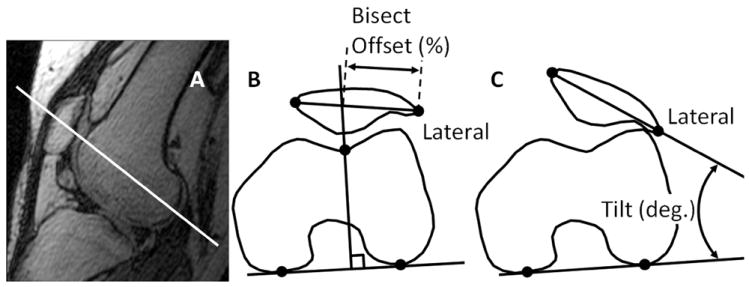
Figure 2.

An oblique-axial plane (solid white line) intersecting the center of the patella and the most posterior points of the femoral condyles was created from 3D MRI volume (A). Anatomical landmarks (black dots) on the oblique-axial plane were used to determine bisect offset (B) and patellar tilt (C).
