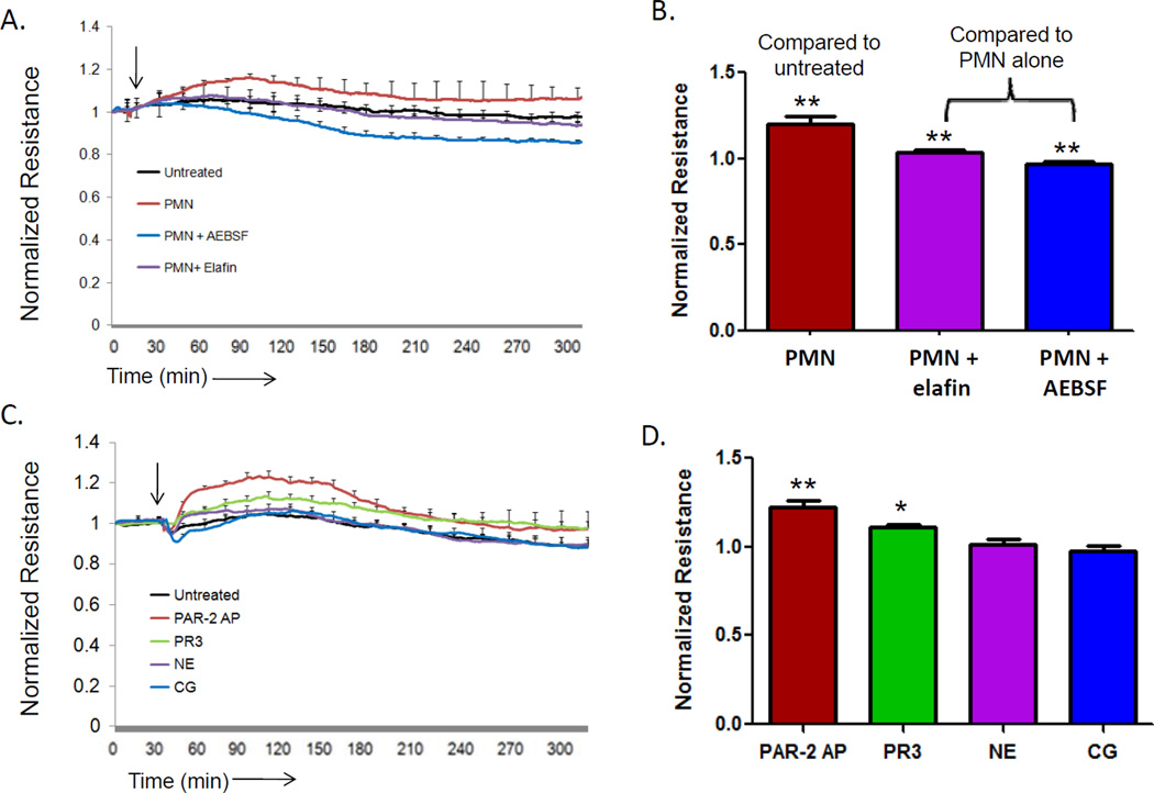
Figure 1. Neutrophil PR3 induces an increase in endothelial cell barrier function.
HUVEC (1x10⁵) cultured on 8W10E+ arrays and barrier function was measured by ECIS. The arrow indicates the time of endothelial cell treatment. (A) HUVEC were stimulated four hours with IL-1β (1 ng/ml) before the addition of neutrophils (1x10⁶) in the presence or absence of serine protease inhibitors AEBSF (10 µM) or elafin 2 µM). Results represent the mean ± SEM of two wells from one of four representative experiments. (B) Neutrophils caused a significant increase in endothelial cell barrier function compared to untreated endothelial cells. Inhibition of neutrophil serine proteases with elafin or AEBSF significantly reduced the increase in endothelial cell barrier function induced by neutrophils (mean ± SEM from four separate experiments, ** p <0.01). (C) Endothelial cells treated with a positive control PAR-2 activating peptide (SLIGKV, 10 µM), proteinase 3 (PR3, 0.1 U/ml), neutrophil elastase (NE, 0.1 U/ml) or cathepsin G (CG, 0.1 U/ml). Results represent the mean ± SEM of two wells from one of four representative experiments. (D) PAR-2 and PR3 induced a significant increase in endothelial cell barrier function 60 minutes after treatment (mean ± SEM from four separate experiments, ** p <0.01).

