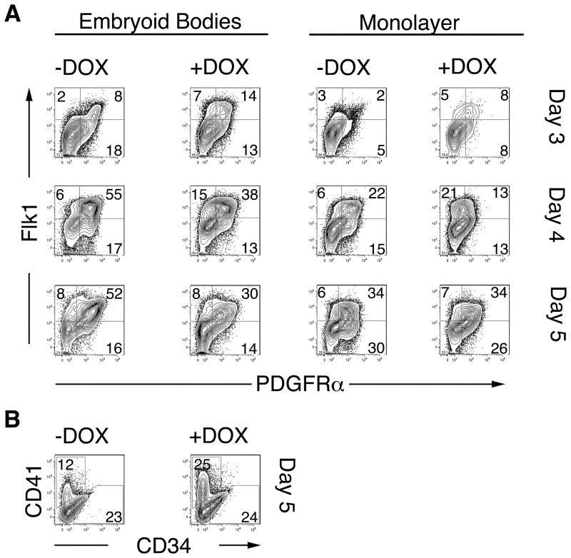
Figure 4. Enhanced mesoderm and hematopoietic development in response to conditional activation of Mixl1 during ES differentiation.
(A) i-Mixl1 ES cells were differentiated as EBs (left panels) or on gelatin-coated dishes as monolayers (right panels) for 3, 4 or 5 days in the presence or absence of DOX. The expression of the mesodermal markers Flk1 and PDGFRα was then examined by flow cytometry. (B) The frequency of CD41+ hematopoietic progenitors and CD34+ vascular endothelial progenitors in EBs cultured in the presence or absence of DOX for 5 days was assessed by FACS. Population frequencies from total, live, gated cells are shown. These data are representative of 3 independent experiments.

