
Figure 1. Calcium transients in sarcoplasmic reticulum and junctional cleft mitochondria.
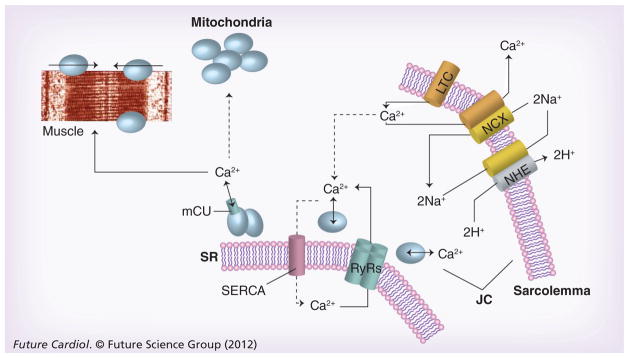
Calcium enters the cardiac myocyte at the beginning of the contraction cycle through LTC. This calcium diffuses through the myocyte to interact with the RyRs on the SR. The SR is a calcium storage organ and stimulation of the RyRs by calcium causes massive calcium release from the SR, which is channeled to muscle fibers where it initiates contraction. At the end of the contraction cycle, calcium is pumped back into the SR by a calcium pump called SERCA. The calcium that originally entered through the LTC is transported back out of the myocyte in exchange for sodium by the NCX exchanger in the sarcolemma. This causes net entry of sodium that can be removed in exchange for protons through the NHE exchanger. Calcium fluxes are highest in the JC between the SR and sarcolemmal membranes, known as a calcium microdomain. Mitochondria are also at high density within the JC and transport calcium in parallel with the calcium transient. Calcium is taken up by mitochondria through an mCU that normally operates in unison with a Na+/Ca2+ antiporter to maintain calcium homeostasis.
JC: Junctional cleft; LTC: L-type calcium channels; mCU: Electrogenic importer; NCX: Sodium calcium exchanger; NHE: Sodium-proton exchanger; RyRs: Ryanodine receptor; SERCA: Sarcoplasmic reticulum calcium ATPase; SR: Sarcoplasmic reticulum.