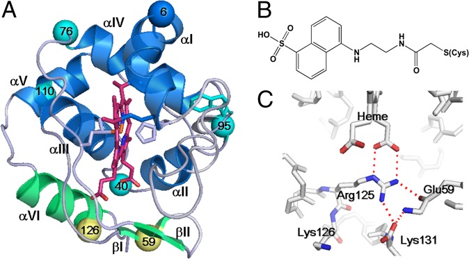
Fig. 1.
(A) Crystal structure of WT c552 obtained at pH 5.44 (PDB ID code 3VNW). Dns-labeled sites are shown as spheres: group I (cyan) and group II (yellow). Helix αVI and β-sheets I and II are distinct structural features of c552 that exhibit group II folding behavior (green). For reference, Trp91 is also highlighted in cyan. (B) Dns-modified cysteine. (C) Red dashed lines indicate electrostatic interactions in the region of distinctive structures of c552.

