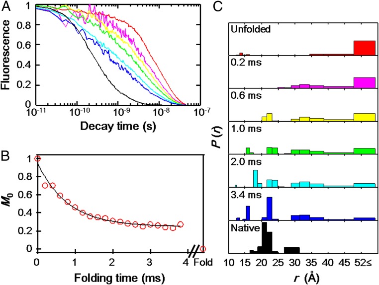
Fig. 4.
Folding kinetics of Dns110–c552 triggered by Gdn jump from 6 to 1 M in a continuous-flow mixer (pH 3.0, ambient temperature). (A) Fluorescence decay curve (7 of the 23 measured decays are displayed): 0 ms (unfolded in 6 M Gdn, red), 0.2 ms (magenta), 0.6 ms (yellow), 1.0 ms (green), 2.0 ms (cyan), 3.4 ms (blue), and >30 min (folded state in 1 M Gdn, black) after the initiation of folding reaction. (B) Integrated Dns fluorescence intensity (M0) as a function of folding time. The solid line is a monoexponential fit with a rate constant of 1,230 s−1. (C) Distributions of P(rDA) for refolding extracted from TR fitting of the fluorescence decay curve (A). For clarity, only 7 of the 23 observed decays are displayed: unfolded in 6 M Gdn (red), 0.2 (magenta), 0.6 (yellow), 1.0 (green), 2.0 (cyan), 3.4 ms (blue), and folded in 1 M Gdn (black).

