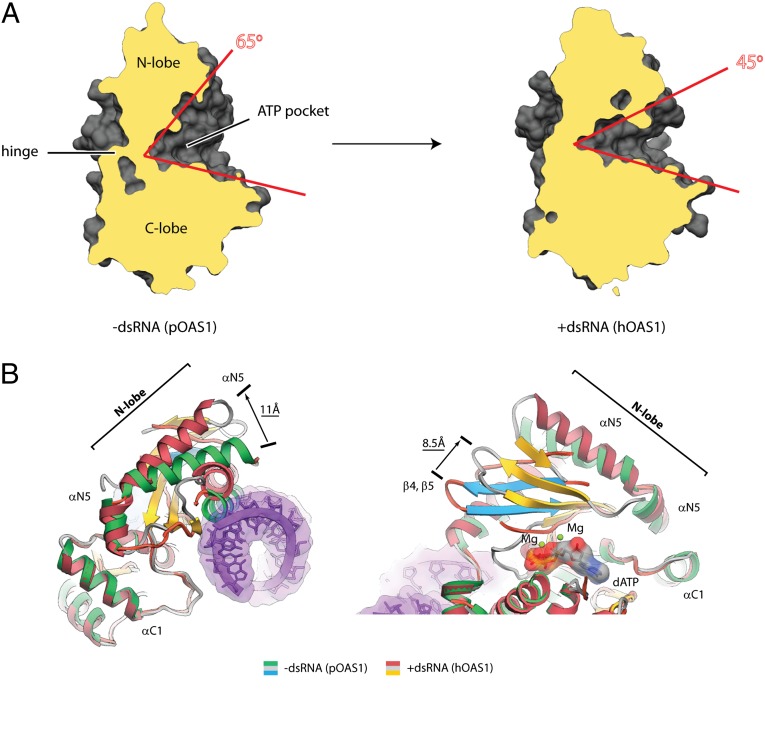
Fig. 3.
Global conformational changes in OAS1 upon dsRNA binding. (A) Side view of the interlobe groove in pOAS1 and in hOAS1. Clipping planes cut the structures at the same position. (B) Superposition of pOAS1 in the inactive conformation (PDB code 1PX5) with hOAS1 in the active conformation (present work). Movement of helix N5 away from the protein/RNA interface and sliding of the β-strand floor that contains the active site residues D75, D77, and D148 are shown.

