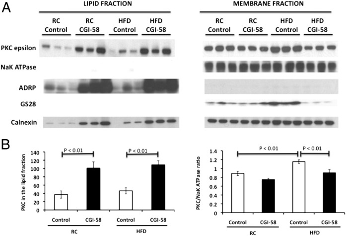
Fig. 6.
PKCɛ translocates to the lipid droplet/ER rather than the membrane with CGI-58 knockdown. (A) Western blot analysis of PKCɛ in the lipid droplet/ER fraction and membrane fraction in mice treated with control or CGI-58 ASO for 8 wk. NaK ATPase (plasma membrane), ADRP (lipid droplet), GS28 (golgi apparatus), and calnexin (ER) (n = 6 per group). (B) Densitometry of the PKCɛ described in A. P value calculated by two-way ANOVA.

