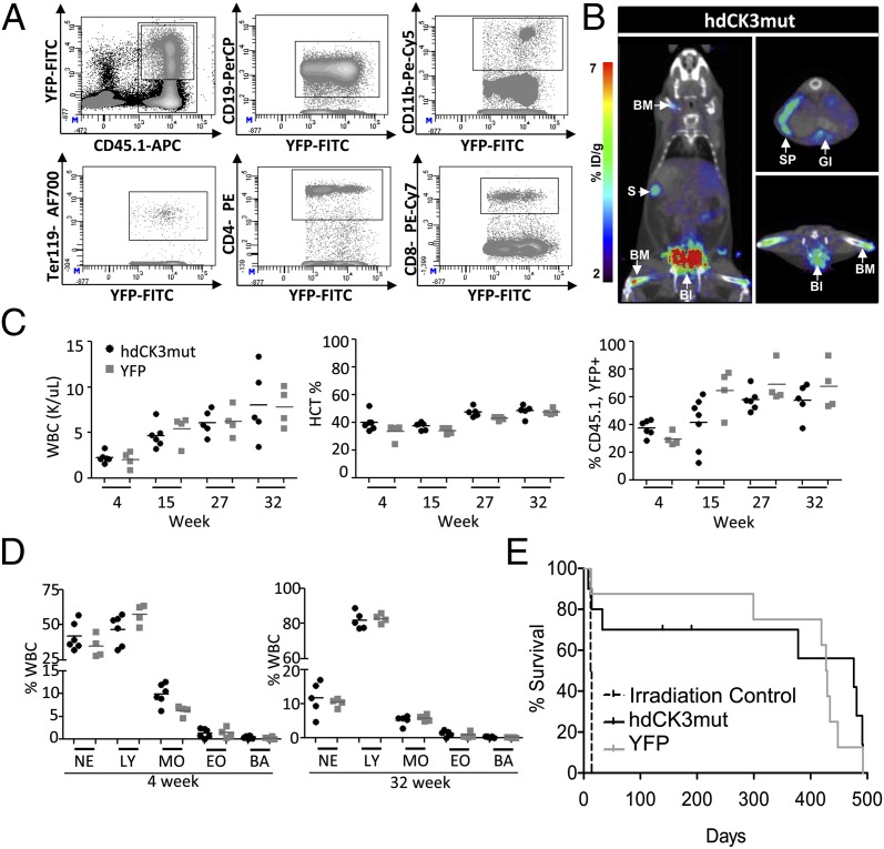
Fig. 3.
hdCK3mut mouse HSCs persist in vivo allowing long-term monitoring of therapeutic cell transplantation. (A) Representative FACS plot for hdCK3mut engraftment within the spleen. Cells were monitored for CD45.1 (donor) and YFP (reporter) positive. Further gating demonstrates that reporter positive (YFP+) cells can be found in all major lineages. (B) [18F]-L-FMAU MicroPET at 32 wk post-BMT. (C) Serial monitoring of peripheral blood. Animals were monitored for total white blood cell (WBC), hematocrit (HCT), and reporter-labeled donor engraftment (CD45.1+, YFP+). (D) Distribution of white blood cells at early and late engraftment are indistinguishable between YFP and hdCK3mut animals. NE, neutrophils; LY, lymphocytes; MO, monocyte; EO, eosinophil; BA, basophil. (E) No survival disadvantage seen in hdCK3mut reporter animals.

