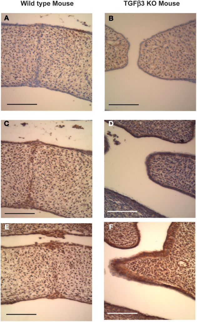
Figure 3.

Coronal sections of C57 Wild-type and TGFβ3 null mouse crania stained for Has expression after palatal shelf elevation (E14.5). Wild-type (A,C and E) and TGFβ3 null (B,D and F) mouse palatal shelves at E14.5 were stained for Has1 using a polyclonal goat anti-Has1 antibody (A), wild-type mouse and (B), TGFβ3 null mouse, Has2 using a polyclonal goat anti-Has2 antibody (C), wild-type mouse and (D), TGFβ3 null mouse or Has3 using a polyclonal rabbit anti-Has3 antibody (E), wild-type mouse and (F), TGFβ3 null mouse. Positive staining appears brown whereas negative staining appears purple. The bars on the diagrams represent 200 μm. The expression of Has1 and 2 appears reduced in the absence of TGFβ3, with the reduction in Has2 being the greatest. Has3 expression may be slightly increased in the absence of TGFβ3.
