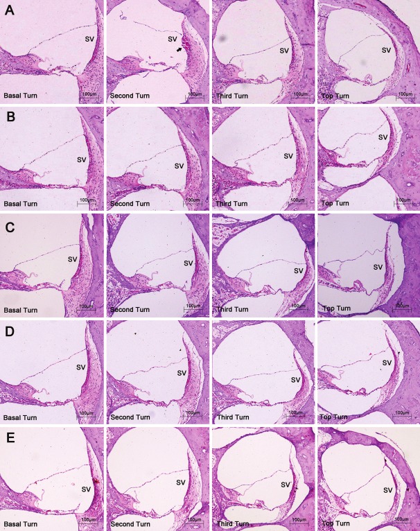
Figure 1.
The pathological changes of the inner ear. Haematoxylin-eosin, original magnification ×100. SV, stria vascularis. A, B, C, D and E: show the pathological changes of the four turns of the cochlea at each distance, respectively. The arrow indicates the rupture of the stria vascularis in the second turn.

