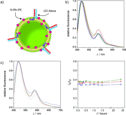
Figure 1.

Study of the stable incorporation of DBCs in liposomes by a FRET assay. a) Illustration of the vesicles constituting the FRET-system: r22-Alexa (donor) is hybridized to c22-b-PPO/DPhyPC liposomes containing N-Rh-PE (acceptor). b) Fluorescence spectra of r22-Alexa/N-Rh-PE pair in FRET (red) and no-FRET (controls, blue and black) systems. Green line shows the spectrum of the FRET system after disrupting the liposomes with Triton X-100. c) Left: Fluorescence spectra of r22-Alexa/N-Rh-PE in the FRET system after mixing with pure DPhyPC liposomes at different v/v ratios: 1:1 (solid green line), 1:10 (solid red line) and 1:100 (solid blue line). Dotted and dashed lines represent the spectra of FRET and no-FRET systems, respectively, as controls without mixing with pristine vesicles. Right: Evolution of I590/I520 over time for the above three ratios.
