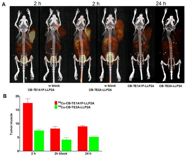
Figure 5.
Small-animal PET/CT imaging of B16F10 tumor-bearing mice at 2 h, with or without coinjection of excess amount of LLP2A and 24 h post-injection of 64Cu-CB-TE1A1P-LLP2A (100 μCi; 0.67 μg) and 64Cu-CB-TE2A-LLP2A (100 μCi; 0.67 μg) (A). The Tumor: background ratios at 2 and 24h were calculated from the MIP images (SUVs) of tumor-bearing C57BL/6 mice (n = 2; bars ± SE) (B).

