
Figure 3.


Donor specific MHC I antibodies increase monocyte recruitment into a murine cardiac allograft.
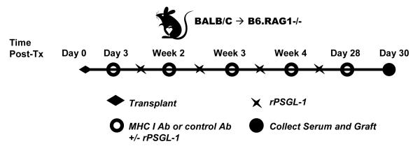
(a) Diagram of antibody administration protocol. BALB/c (H-2d) donor hearts were heterotopically grafted into RAG1 knockout recipients on a C57BL/6 background (H-2b). Animals were passively transferred with a murine isotype control monoclonal IgG2a or monoclonal directed against the donor MHC I locus beginning on day 3 after transplant and continuing weekly thereafter until day 28. Grafts were recovered from recipients while still beating on day 30 post-transplant. Cellular infiltration in the base was assessed by immunohistochemical staining and scored by a pathologist blinded to the experimental groups. The fusion protein rPSGL-1-Ig was administered to a group of animals biweekly beginning on day 3 post-transplant at 70μg per animal.
(b) The presence of circulating donor specific MHC I antibodies in recipient serum was confirmed using flow cytometric crossmatch on BALB/c splenocytes. BALB/c splenocytes were incubated with 25μL of neat serum collected from allograft recipients, and stained with anti-mouse Fcγ-FITC. Cells were stained with anti-CD3-PE and fluorescence was measured by flow cytometry. CD3-positive cells were gated as shown in the representative dot plot, and the median fluorescence in the FL-1 channel was determined. Results are shown as median fluorescence intensity of serum on CD3 positive cells +/−SEM, using sera from multiple animals for each condition: unstained (n=5), control mIgG2a (n=3), anti-Kd mIgG2a (n=7), anti-Dd mIgG2a (n=2), anti-Kd + Dd mIgG2a (n=2).
(c) Native or transplanted hearts were assessed immunohistochemically for Mac-2 and staining was scored by a pathologist blinded to the experimental groups. The box and whiskers graph shows the distribution of scores for each group. Infiltration near the suture site or in the endocardium was disregarded. Global ANOVA, then individual groups. * p<0.05, ** p<0.01, *** p<0.001 versus control mIgG2a group.