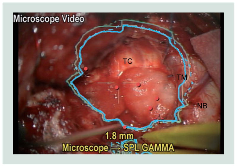
Figure 4. Injection of navigational information into microscopic view.

In this view generated by the neuronavigational system, an image of the microscope video feed in the bottom left-hand corner demonstrates the surgeon’s view of the outline (blue) of the preoperatively segmented lesion that is being resected.
NB: Normal brain adjacent to the operative cavity; TC: Tumor cavity; TM: Tumor margin.
Colour figure can be found online at www.expert-reviews.com/doi/full/10.1586/erd.12.42
