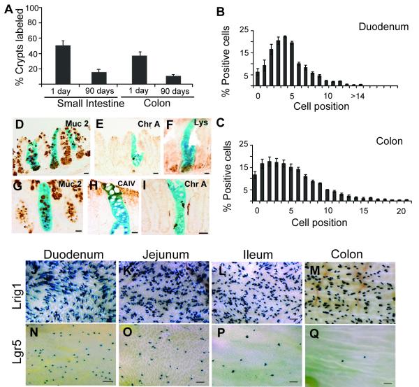
Figure 2. β-gal labels Lrig1+ cells in the SC niche, persists in the long-term and labeling differs in Lgr5-reporter mice.
(A) One day after tamoxifen induction, 50% of small intestinal crypts and 40% of colonic crypts are labeled. At 90 days, the number of labeled crypts decreases to 18% in the small intestine and 10% in the colon. (B-C) Frequency of β-gal+ cells at different positions relative to the small intestinal (B) and colonic (C) crypt base one day after tamoxifen administration. (D-I) Co-staining with various differentiation markers to confirm multipotency of progeny of β-gal+ cells: Mucin2 (Muc 2) marks goblet cells (D,G); Chromogranin-A (Chr A) marks enteroendocrine cells (E,I); Lysozyme (Lys) marks Paneth cells (F); and Carbonic Anhydrase IV (CAIV) marks enterocytes (H). (J-M) Whole-mount view of Lrig1-CreERT2/+;R26RLacZ/+ and (N-Q) Lgr5-EGFP-IRES-CreERT2;R26RLacZ/+ lineage-labeled intestines. Error bars represent s.e.m. Scale bars in D-I represent 25μm and J-Q represent 500μm. See also Figure S2.

