
Figure 7. Homozygous Lrig1-CreERT2 mice exhibit up-regulation of ErbB1-3 and develop duodenal adenomas and superficially invasive carcinomas.
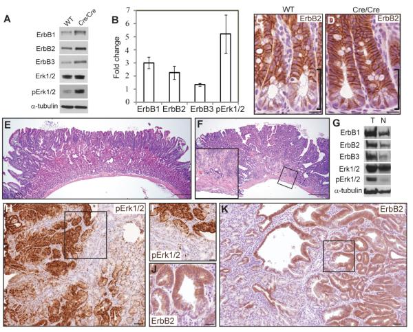
(A) Representative western blot showing increased ErbB1-3 and pErk1/2 levels in crypt epithelium isolated from small intestine of Lrig1-CreERT2/CreERT2 (Cre/Cre) mice compared to wild-type (WT) mice. (B) Quantification of protein expression levels shown in (A). (C-D) Immunohistochemical examination of ErbB2 (brown) in intestinal crypts from WT (C) and Cre/Cre (D) mice. Black brackets indicate differential staining at the crypt base. (E) Representative H&E staining of an adenoma from a five-month-old Lrig1-CreERT2/CreERT2 mouse. Tumors exhibited low-grade dysplasia and a marked plaque-like expansion of Brunner’s glands (n=14). (F) One tumor exhibiting histological progression: areas of cribiform architecture and loss of cellular polarity and focal extension of neoplastic glands into deeper layers of the bowel wall, suggestive of early invasion (inset). (G) Representative western blot showing increased ErbB1-3 and pErk1/2 levels in tumor (T) compared to gross normal duodenum (N) of Lrig1-CreERT2/CreERT2 mice. (H-I) Immunohistochemical examination of pErk1/2 (brown in H) in tumors from Lrig1-CreERT2/CreERT2 mice; boxed region magnified in I. (J-K) Immunohistochemical examination of ErbB2 (brown in K) in tumors from Lrig1-CreERT2/CreERT2 mice; boxed region magnified in J. Scale bars represent 25μm in C, D, I and J, represent 100μm in E and F and 75μm in (H and K). Error bars represent s.e.m. See also Figure S7 and Table S4.