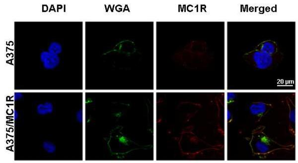
Figure 1.
ICC of MC1R expression on the surface of A375 parental and engineered cells. Confocal micrographs of cells incubated with the nuclear marker DAPI (blue), the plasma- and plasma-membrane marker, WGA (green) and MC1R antibody-Alexa 555 (red). To inhibit cellular uptake, cells were incubated with antibodies and dyes at 4°C for 10 min. The merged image shows co-localization of MC1R (red) with membrane marker (green) indicating accumulation of the receptor on the cell-surface (yellow).

