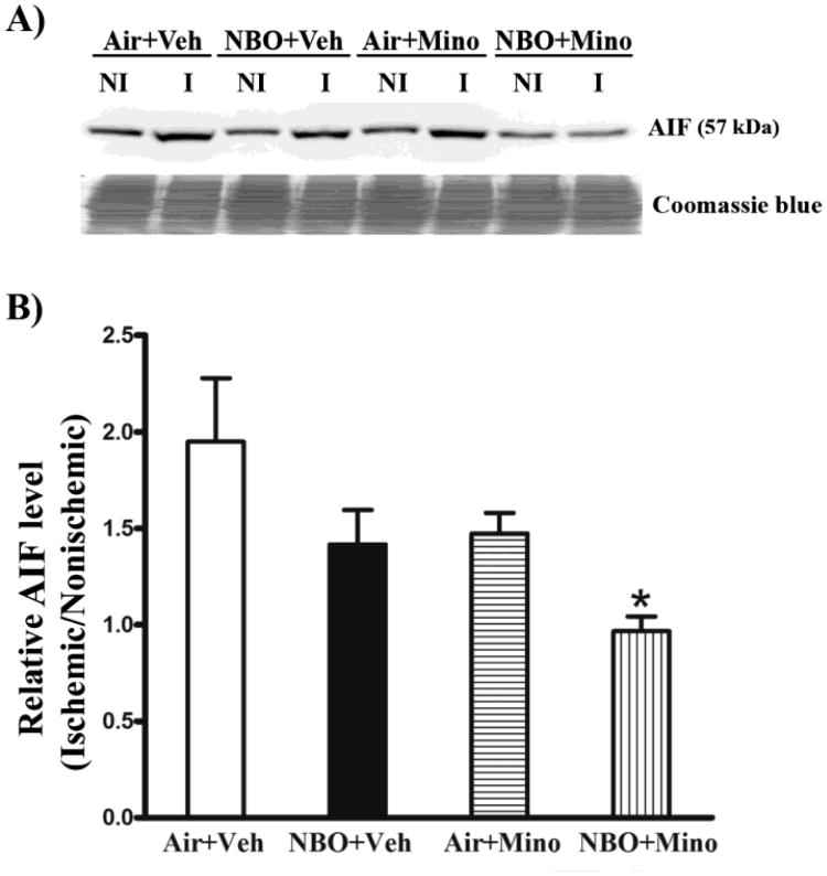
Fig. 5.
Effects of NBO, minocycline and their combination on AIF induction in the ischemic brain after 90-min MCAO and 48 hrs of reperfusion. Western blot was conducted to detect AIF protein in the nonischemic (NI) and ischemic (I) hemispheric tissue. Veh: vehicle; Mino: minocycline. (A) A representative western blot revealed AIF changes in the ischemic brain of each group (upper panel). The membrane was stained with Coomassie blue as a loading control (lower panel). (B) AIF protein was quantitated after normalization to the intensity of Coomassie staining and expressed as hemispheric ratio (ischemic/nonischemic). Cerebral ischemia and reperfusion induced a 0.94-fold increase in AIF protein in the ischemic tissue, which was almost completely inhibited by the combination therapy (*P < 0.05 versus Air + Veh). NBO or minocycline alone resulted in a small but not significant reduction in AIF increase. All three treatments did not affect AIF level in the nonischemic hemisphere. Data are expressed as mean ± SEM, n = 8 for each group.

