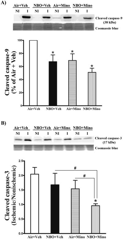
Fig. 6.
Effects of NBO, minocycline and their combination on caspase-3 and -9 cleavage in the ischemic brain after 90-min MCAO and 48 hrs of reperfusion. Western blot was conducted to detect cleaved caspase-3 and -9 in the nonischemic (NI) and ischemic (I) hemispheric tissue. Veh: vehicle; Mino: minocycline. (A) The combination therapy led to a greater inhibition on caspase-9 cleavage than NBO or minocycline alone. A representative western blot showed that cleaved caspase-9 was barely seen in nonsichemic (NI) hemispheric tissue, but was significantly increased in ischemic (I) tissue (Upper panel). After normalization to the total intensity of Coomassie staining (Middle panel), cleaved caspase-9 was quantitated and expressed as a percentage of AIF level in the ischemic hemispheric tissue of the Air + Veh group (Bottom panel). NBO, minocycline or their combination significantly reduced the level of cleaved caspase-9 in ischemic tissue (*P < 0.05 versus Air + Veh), and a greater reduction was seen for the combination therapy (#P < 0.05 versus NBO + Veh or Air + Mino). (B) The combination therapy, but not NBO or minocycline alone, significantly reduced caspase-3 cleavage in ischemic hemispheric tissue. A representative western blot showed cleaved caspase-3 levels in the nonischemic and ischemic hemispheric tissue of each group (Upper panel). After normalizing to Coomassie staining (Middle panel), cleaved caspase-3 was quantitated and expressed as a hemispheric ratio (ischemic/nonischemic) (Bottom panel). Data are expressed as mean ± SEM. *P < 0.05 versus Air + Veh, n = 8 for each group.

