
Fig. 3.
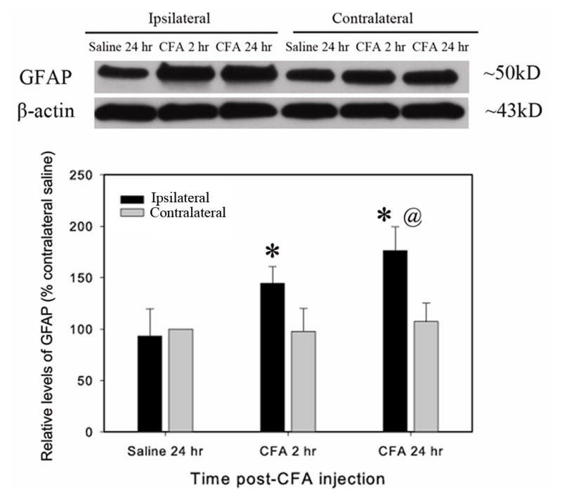
Spinal GFAP expression during CFA-induced peripheral inflammation. CFA was subcutaneously injected into the hind paw. A. A representative GFAP western blot. Note that GFAP was significantly upregulated in the ipsilateral but not the contralateral spinal dorsal horn. B. Quantification of relative levels of GFAP. The values were normalized using saline-injected rats as control (100%). CFA induced a significant increase 2–24 h post CFA-injection compared to saline-injected control. *P<0.05 vs. saline-injected control and @P<0.05 vs. contralateral side (n=4 per group).