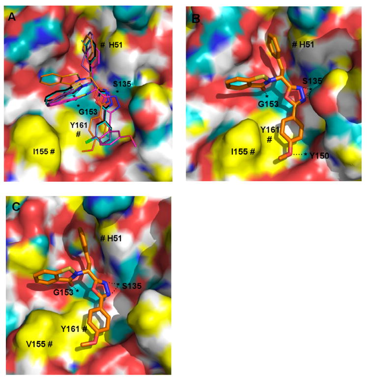
Figure 6. Putative binding mode of selected compounds interacting with DENV2 (A and B) and WNV (C) proteases, as predicted by molecular modeling.

All compounds have CPK-colored heteroatoms, and are distinguished according to the color of their carbon atoms as follows: black = 7i, cyan = 7w, purple = 7x, magenta = 7g and orange = 7n. The protease receptor surfaces are colored as follows: yellow = lipophilic, white = weakly polar, cyan = polar H, blue = polar N and red = polar O. Apparent H-bonding features of the receptor are marked with “*” and hydrophobic interactions with “#”
