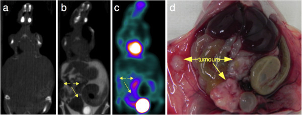
Figure 6.

Impact of intraperitoneal and intravenous contrast media enhancement on tumor localization on SA-CT images: mice. Representative coronal slices for unenhanced CT (a), contrast-enhanced SA-PET/CT (b and c), and necropsy photograph of a mouse with multiple abdominal lesions and hemorrhagic ascites (d). The animal first underwent SA-CT acquisition, was subsequently injected with a mixture of 18F-FDG and Fenestra VC, and received an intraperitoneal injection of iohexol immediately before the SA-PET/CT acquisition began. Tumors are well defined on the contrast-enhanced CT slice (yellow arrows), including a necrotic lesion located near the bladder harboring a low 18F-FDG uptake and a central photopenic area on an SA-PET slice. Also visible is a tumor at the site of tumor cell injection.
