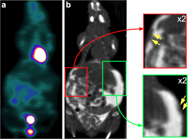
Figure 7.
Example of carcinomatosis lesions depicted by contrast-enhanced CT and overlooked by PET. Representative coronal slices for 18F-FDG SA-PET/CT (a) and contrast-enhanced CT (b) of a mouse with multiple abdominal lesions. Small carcinomatosis lesions (insets, yellow arrows) involving the abdominal wall are visible on CT but are not 18F-FDG avid.

