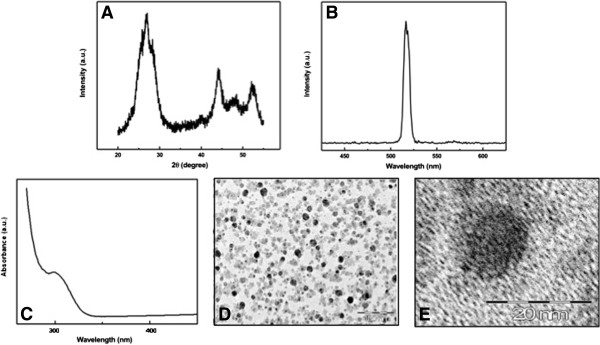
Figure 1.

Quantum dot particles’ formation and characterization. (A) X-ray diffraction patterns of CdS-MD nanoparticles. (B) Emission profiles of CdS-MD nanoparticles. (C) UV-visible spectrum of CdS-MD nanoparticles. (D and E) TEM images of CdS-MD nanoparticles. The particles appear evenly spread in the polymeric matrix of maltodextrin. Although some clusters of two to four QDs are visible, most of the QDs are isolated, suggesting that the majority are monodispersed single QD.
