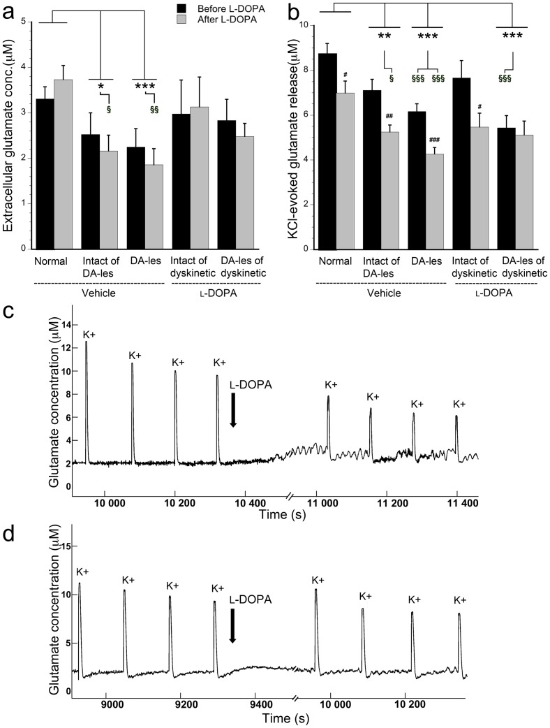
Figure 2. Quantification (in µM) of basal extracellular glutamate concentration (a) and KCl-evoked glutamate release (b) before and after acute l-DOPA administration in normal rats and in bilateral striata of vehicle- and l-DOPA-treated animals.
Extracellular glutamate concentration was estimated by averaging the baseline and glutamate release was observed upon KCl-ejections (K+) during in vivo recordings, here in normal striatum (c) and dopamine-lesioned striatum of a dyskinetic subject (d). #p<0.05, ## p<0.01, and ###p<0.001 compared to before acute l-DOPA administration in the same group of animals. * p<0.05, ** p<0.01, and *** p<0.001 for comparisons between groups with two-factor ANOVAs and § p<0.05, §§ p<0.01, and §§§ p<0.001 for comparisons between groups with one-way ANOVA. Dopamine-lesioned striata is abbreviated DA-les.

